Diagnosis of Melanoma
It is important to be aware that melanomas can occur anywhere on the skin surface, but they most frequently develop on the back, arms, neck, scalp and legs. It is therefore important that the entire skin surface is examined when screening for melanomas.
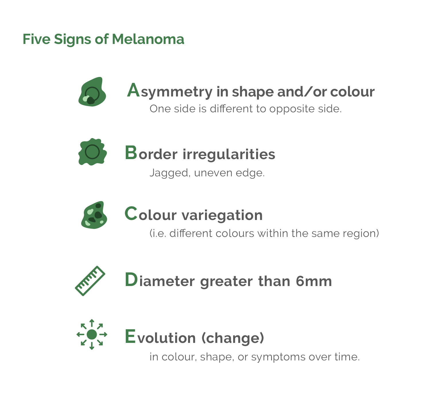
Five Signs of Melanoma
Ultimately, melanomas are diagnosed by microscopic examination of a biopsy specimen by a pathologist. Biopsying the correct mole or freckle requires one to know what the suspicious features of a melanoma are. The features of a mole/freckle that would make one suspicious of a melanoma can best be remembered by using the acronym ABCDE (see below). If it is not possible to remember this then remember to look out for new funny-looking freckles that are enlarging.
• A : Asymmetry (one side is different to opposite side)
• B : Border irregularities (jagged, uneven edge)
• C : Colour variegation (i.e, different colours within the same region)
• D : Diameter greater than 6 mm
• E : Evolution (change) in colour, shape, or symptoms over time
Other abnormal features include inflammation (redness of the skin around the freckle), bleeding and crusting (scabbing). Moles/freckles that are inflamed or crusting are often slightly sensitive and people describe the freckle as having an unpleasant itch. A person who notices any of these changes should make an appointment with their doctor as soon as possible.
The most dangerous melanomas are ones that appear suddenly and grow rapidly because they have a high risk of metastasising early on. They can have very bizarre appearances and look nothing like a typical melanoma. So, any new rapidly growing spot on the skin ought to be examined promptly by a professional because it could be a melanoma.
Mole Mapping for Melanoma
Specialists make use of skin microscopes called dermatoscopes to help improve the accuracy of clinical diagnosis of melanomas. Magnification of the mole or freckle allows a far better and more complete analysis which reduces the number of spots that are biopsied unnecessarily. Mole mapping takes this one step further and allows one to take magnified digital pictures of moles. These pictures are stored and can be compared with pictures taken of the same mole some months later. It is thus possible to detect subtle and early changes in the mole and improve one's ability to diagnose an early melanoma. Mole mapping is indicated when people have multiple suspicious looking moles, not just a few normal freckles.
The Four Main Types of Melanoma
It is important to note that the type of melanoma that one has does not influence prognosis.
Most melanomas (90%) occur as new growths on the skin.
Only 10% of melanomas grow from existing moles or freckles. So when monitoring one’s skin, one should preferentially be looking for new freckles that are enlarging. This explains why it does not make sense to remove all of a person’s moles to try and prevent a melanoma from occurring because most of the time the melanoma will be a new growth.
Click here to see pictures of the different types of melanomas.
1. Superficial Spreading Melanoma
By far the most common (70%) is called superficial spreading melanoma and as the name implies, it starts on the surface of the skin (in the epidermis) and enlarges. It generally takes the appearance of a funny-looking freckle which is flat, irregular in shape and has various colours ranging from pink and brown to black. It is important to note that the most common form of melanoma starts as a totally flat growth.
2. Nodular Melanoma
The common perception is that a melanoma forms a bump on the surface of the skin, but this only happens 5% of the time. Melanomas that form bumps are known as nodular melanomas. They generally grow faster than the superficial spreading variety and penetrate into the skin at an earlier stage of their development.
3. Lentigo Maligna Melanoma
Lentigo maligna melanoma occurs when a lentigo maligna turns cancerous. Lentigo malignas are the smudgy brown sun spots that commonly develop on the cheeks of elderly people. Change within a brown mark on the face (in particular, becoming darker) could signal the beginnings of a melanoma and a biopsy is indicated.
4. Acral Lentiginous Melanoma
This is a form of melanoma that occurs on the peripheries (acral is Greek for peripheral) such as one's hands and feet. The classical acral lentiginous melanoma is one that occurs under the nail and can look just like a bruise under the nail.
Amelanotic Melanoma
Special mention needs to be made about amelanotic melanomas because they are frequently missed and often diagnosed too late. They are not considered a separate category of melanoma and can be classified as superficial spreading, nodular or acral. Amelanotic melanomas do not produce any pigment at all and appear as pink or reddish spots which can be totally flat or produce bumps. They are rare, accounting for only 5% of all melanomas and tend to appear and grow rapidly (over weeks to a few months).
What is a Biopsy for a Melanoma?
If a suspicious mole is discovered it needs to be excised (biopsied) and sent to the pathology laboratory for analysis. Usually the entire spot is removed under local anaesthetic in the doctor's rooms (if too large to remove without a skin graft or flap, then the most sinister-looking area of the mole is removed) and examined under a microscope by a pathologist to determine if precancerous or cancerous cells are present.